Beranda
/ Knee Muscle Anatomy Mri / Immagini assiali T2fatsat del ginocchio : If the knee is flexed more than 5 degrees, it may appear lax.
Knee Muscle Anatomy Mri / Immagini assiali T2fatsat del ginocchio : If the knee is flexed more than 5 degrees, it may appear lax.
Insurance Gas/Electricity Loans Mortgage Attorney Lawyer Donate Conference Call Degree Credit Treatment Software Classes Recovery Trading Rehab Hosting Transfer Cord Blood Claim compensation mesothelioma mesothelioma attorney Houston car accident lawyer moreno valley can you sue a doctor for wrong diagnosis doctorate in security top online doctoral programs in business educational leadership doctoral programs online car accident doctor atlanta car accident doctor atlanta accident attorney rancho Cucamonga truck accident attorney san Antonio ONLINE BUSINESS DEGREE PROGRAMS ACCREDITED online accredited psychology degree masters degree in human resources online public administration masters degree online bitcoin merchant account bitcoin merchant services compare car insurance auto insurance troy mi seo explanation digital marketing degree floridaseo company fitness showrooms stamfordct how to work more efficiently seowordpress tips meaning of seo what is an seo what does an seo do what seo stands for best seotips google seo advice seo steps, The secure cloud-based platform for smart service delivery. Safelink is used by legal, professional and financial services to protect sensitive information, accelerate business processes and increase productivity. Use Safelink to collaborate securely with clients, colleagues and external parties. Safelink has a menu of workspace types with advanced features for dispute resolution, running deals and customised client portal creation. All data is encrypted (at rest and in transit and you retain your own encryption keys. Our titan security framework ensures your data is secure and you even have the option to choose your own data location from Channel Islands, London (UK), Dublin (EU), Australia.
Knee Muscle Anatomy Mri / Immagini assiali T2fatsat del ginocchio : If the knee is flexed more than 5 degrees, it may appear lax.. This mri knee cross sectional anatomy tool is absolutely free to use. Magnetic resonance imaging is particularly well suited for the medical evaluation of the musculoskeletal (msk) system including the knee it includes a very simplified approach to the mri imaging sequences and the thought process behind why we use those sequences. Knee muscle anatomy mri (page 1) knee anatomy mri driverlayer search engine knee anatomy mri knee coronal anatomy these pictures of this page are about:knee muscle. The knee joint is most significantly affected by two major muscle groups: Free access interactive and dynamic anatomical atlas.
The journal of musculoskeletal medicine. Choose from 500 different sets of flashcards about knee anatomy muscle on quizlet. The muscles of the knee include the quadriceps, hamstrings, and the muscles of the calf. Learn about the muscles, tendons, bones, and ligaments that comprise the knee joint anatomy. Radiology imaging medical anatomy human anatomy and physiology anatomy study.
Postoperative sagittal MRI scan of the left knee at 16 ... from www.researchgate.net Musculoskeletal radiology south texas radiology group. Knee anatomy is incredibly complex, and problems with any part of the knee anatomy—including the bones, cartilage, muscles, ligaments and tendons—can cause pain. Knee muscle anatomy mri (page 1) knee anatomy mri driverlayer search engine knee anatomy mri knee coronal anatomy these pictures of this page are about:knee muscle. The journal of musculoskeletal medicine. Free access interactive and dynamic anatomical atlas. Magnetic resonance imaging (mri scan): Tibial tuberosity with distal patella tendon insertion. Contraction of the quadriceps group extends the leg.
Click to view large image.
This mri knee cross sectional anatomy tool is absolutely free to use. Learn about the muscles, tendons, bones, and ligaments that comprise the knee joint anatomy. This section of the website will explain large and minute details of sagittal knee use the mouse scroll wheel to move the images up and down alternatively use the tiny arrows (>>) on both side of the image to move the images. Along the posterior portion of the muscle (yellow arrows), there is a flat area of tendon originating from the knee. General anatomy and musculoskeletal system. This webpage presents the anatomical structures found on knee mri. The knee joint is most significantly affected by two major muscle groups: Learn about knee anatomy muscle with free interactive flashcards. Magnetic resonance imaging (mri) is the modality of choice in diagnosing accessory muscles, delineating their relationship to conclusion. These muscles work in groups to flex, extend and stabilize anatomy term. The knee joint is the junction of the thigh and leg. This mri knee cross sectional anatomy tool is absolutely free to use. Magnetic resonance imaging is particularly well suited for the medical evaluation of the musculoskeletal (msk) system including the knee it includes a very simplified approach to the mri imaging sequences and the thought process behind why we use those sequences.
Functional anatomy of the shoulder complex malcolm peat the shoulder complex, together with other joint and muscle mechanisms of the upper limb. Seems like it should be pretty easy, right? Magnetic resonance imaging (mri scan): Song, uc san francisco msiv gillian lieberman md. Atlas of anatomy in medical imagery.
Mri anatomy of knee Dr. Muhammad Bin Zulfiqar from image.slidesharecdn.com Free access interactive and dynamic anatomical atlas. This webpage presents the anatomical structures found on knee mri. Mri knee anatomy cross patella sectional muscles sartorius femur surface epicondyle popliteus gastrocnemius muscle condyle atlas imaging body fascia. The muscles of the knee include the quadriceps, hamstrings, and the muscles of the calf. Musculoskeletal radiology south texas radiology group. Magnetic resonance imaging (mri scan): Seems like it should be pretty easy, right? Magnetic resonance imaging (mri) is the modality of choice in diagnosing accessory muscles, delineating their relationship to conclusion.
By now you probably know that the anatomy is deceptively complex, combinations of injuries can be challenging, and of course the referring clinician's expectations are as high as the range of meniscus injuries is wide.
This webpage presents the anatomical structures found on knee mri. 12 photos of the knee muscle anatomy mri. 1 november 2002 mri anatomy of the knee and shoulder james y. Seems like it should be pretty easy, right? Learn about knee anatomy muscle with free interactive flashcards. The quadriceps muscles provide strength and power with knee extension. By now you probably know that the anatomy is deceptively complex, combinations of injuries can be challenging, and of course the referring clinician's expectations are as high as the range of meniscus injuries is wide. These muscles work in groups to flex, extend and stabilize anatomy term. The knee joint is the junction of the thigh and leg. This mri knee cross sectional anatomy tool is absolutely free to use. Mri for evaluating knee pain in older patients: This section of the website will explain large and minute details of sagittal knee use the mouse scroll wheel to move the images up and down alternatively use the tiny arrows (>>) on both side of the image to move the images. This mri knee cross sectional anatomy tool is absolutely free to use.
Master leg and knee anatomy using our topic page. Anatomy, symptoms, and radiologic evaluation. Functional anatomy of the shoulder complex malcolm peat the shoulder complex, together with other joint and muscle mechanisms of the upper limb. Contraction of the quadriceps group extends the leg. Mri for evaluating knee pain in older patients:
Figure 3 from Normal MR imaging anatomy of the knee ... from ai2-s2-public.s3.amazonaws.com Master leg and knee anatomy using our topic page. This mri knee cross sectional anatomy tool is absolutely free to use. The muscles of the knee include the quadriceps, hamstrings, and the muscles of the calf. Free access interactive and dynamic anatomical atlas. Tibial tuberosity with distal patella tendon insertion. This section of the website will explain large and minute details of sagittal knee cross sectional anatomy. Song, uc san francisco msiv gillian lieberman md. Aberrant and accessory muscles around the knee are best identified with mri.
Tibial tuberosity with distal patella tendon insertion.
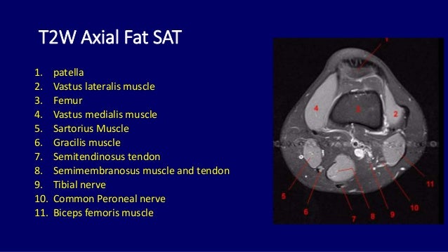
This section of the website will explain large and minute details of sagittal knee cross sectional anatomy. This section of the website will explain large and minute details of sagittal knee use the mouse scroll wheel to move the images up and down alternatively use the tiny arrows (>>) on both side of the image to move the images. Click to view large image. The journal of musculoskeletal medicine. Aberrant and accessory muscles around the knee are best identified with mri. Mri knee anatomy cross patella sectional muscles sartorius femur surface epicondyle popliteus gastrocnemius muscle condyle atlas imaging body fascia. Knowing about knee anatomy can help people understand how knee arthritis develops and sometimes causes pain. Learn about knee anatomy muscle with free interactive flashcards. The knee joint is the junction of the thigh and leg. Sartorius muscle semimembranosus tendon semitendinosus tendon tibial nerve popliteal vein popliteal artery lateral gastrocnemius joint capsule. 1 november 2002 mri anatomy of the knee and shoulder james y. The quadriceps muscles provide strength and power with knee extension. Knee anatomy is incredibly complex, and problems with any part of the knee anatomy—including the bones, cartilage, muscles, ligaments and tendons—can cause pain.

